Motivation: Breast cancer is the most common type of cancer in women and one of theleading causes of female mortality worldwide. There is a need to develop newer andbetter diagnostic tools for the detection of breast cancer and bio-impedance is hypothesizedas one such approach. The utility of this technology originates from the fact that impedancecan capture and reflect the rich biological and electrical characteristics exhibited at the wholebody, organ, tissue, cell and organelle level. The recent development of micro/nanotechnology has provided additional advantages and introduces new paradigms into the investigation of traditional electrical impedance concepts. Miniaturized sensors have significant advantagessuch as high throughput, small characteristic dimensions, low power consumption, andportability. While bio-impedancemeasurements for cells has been studied previously, the advantage of characterizingmicro-scale tissues over cells is that the degree of malignancy as well as the architecturalchanges that occur during the progression of cancer can be quantified on a larger micro-scalesample to make a deterministic assessment compared to studying a single cell. We hypothesize that MEMS based microchip having interdigital electrodes (IDEs) could be used to quantify the electrical properties of the benign and cancerous breast tissue. We use breast tissue cores placed on the IDEs using Tissue Microarray Technology (TMA). The salient features of this project comprise: (a) development of robust and low cost microchips having IDES inside SU-8 well and (b) a high throughput electrical characterization of benign and cancerous breast tissues using a semi-automated bio-impedance measurement system.
Project Highlights:
Fabrication of Microchips with Interdigital Electrodes inside SU-8 Well and Placing Individual Breast Tissue Cores on IDEs
Inter-digitated electrodes(IDEs) are implemented in various sensing devices such as surface acoustic wave (SAW)sensors, chemical sensors as well as MEMS based biosensors. However, while IDEshave been optimized for a variety of sensing applications, the use of IDEs for accessingcancer tissue samples has yet to be adequately investigated. We have fabricated microchip integrated with IDEs for measuring electrical properties of the benign and cancerous breast tissue. To make the sensor biocompatible, the sensor was fabricated on 1.0 mm-thick glass substrate(Pyrex 7740). The microchip was fabricated using a two-mask process. The fabricationprocess is shown in Fig. 1.

Fig. 1. Process flow for the microchip.
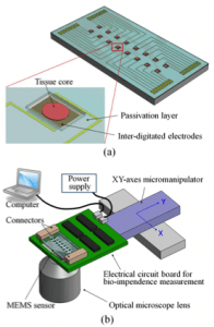
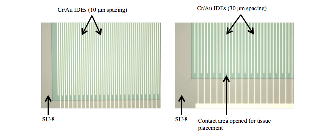
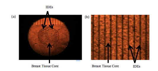
After cleaning glass using a 70:30 solution of H2SO4 and H2O2[Fig. 1(a)], Cr/Au (0.1 μm/0.5 μm) was deposited using thermal evaporation on glasssubstrates [Fig. 1(b)]. Using a standard photolithography technique interdigital electrodes (10μm width with 10 μm spacing and 30 μm spacing) were patterned [Fig. 1(c)]. SU-8photoresist (7 μm thick) was spin coated on glass substrate [Fig. 1(d)] and was patternedusing photolithography to form reservoir [Fig. 1(e)]. The reservoir is made to hold the tissueand medium inside the desired area. The schematics of (a) the MEMS-based bio-impedance sensor for tissue microarray, and (b) the automated bio-impedance measurement system is shown in Fig. 2. Figure 3(a) and 3(b) shows the opticalphotographs of the fabricated microchips with 10 μm and 30 μm spacing, respectively. The Tissue core was prepared using standard Tissue Micro Array (TMA) technique. Individual tissue cores were sectioned at 4 μm in thickness and carefully positioned in the center of the interdigited electrodes. The microchips were deparaffinized: Xylene (5 min, 3 times); 100% Alcohol (5 min, 2 times); 95% Alcohol (5 min, 1 time); 75% Alcohol (5 min, 1 time); rinse in PBS (1-2 times) and immersed in PBS holding solution until impedance analysis. Fig. 4 shows the optical photograph of cancerous tissue placed on the interdigital electrodes having 30 μm spacing.
Fig. 2. The schematics of (a) MEMS-based bio-impedance sensor for tissue microarray and (b) automated bio-impedance measurement system.
Fig. 3.Optical photographs of (a) microchip with IDEs having 10 μm width and 10 μm spacing and (b) 10 μm width and 30 μm spacing.
Fig. 4. Optical photograph of cancerous tissue placed on interdigital electrodes: (a) 4x and (b) 20x magnification.
Bio-Impedance Measurements of Benign and Cancerous Breast Tissue
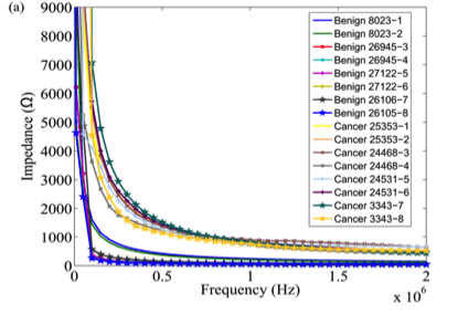
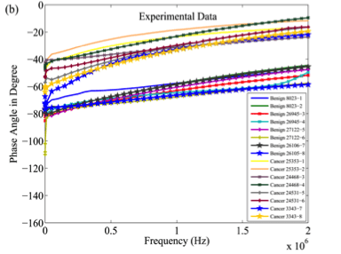
Impedance measurements were performed for the benign and cancerous breast tissues placed on IDEs using an Agilent E4980A impedance analyzer. Eight different specimens with two cores from each specimen (4 benign) and (4 cancerous) were used for impedance measurement. Microchip having IDEs with 10 μm spacing was used for a total of 16 measurements. As seen from Fig. 5, benign and cancerous breast tissues have significant differences in their impedance and phase, which can be identified using microchip and impedance analyzer.In our future work, we plan to integrate bio-impedance measurement with mechanical characterization for automated sampling of breast tissue core specimens.
Fig.9. Plots of (a) impedance and (b) phase measurement for benign and cancerous breast tissues using microchip having IDEs with 10 μm spacing.
Personnel: Hardik J. Pandya, Rajarshi Roy
Sponsor: NIH Grant R01CA161375
Acknowledgment: Research reported in this publication was supported by the National Cancer Institute of the National Institutes of Health under Award Number R01CA161375. The content is solely the responsibility of the authors and does not necessarily represent the official views of the National Institutes of Health.
Publications:
- Hardik J. Pandya, Hyun Tae Kim, Rajarshi Roy, Wenjin Chen, Lei Cong, Hua Zhong, David Foran, and Jaydev P. Desai, “Towards an Automated MEMS-based Characterization ofBenign and Cancerous Breast Tissue using Bio-Impedance,” Sensors and Actuators B, vol. 19, pp. 163-173, 2014.
